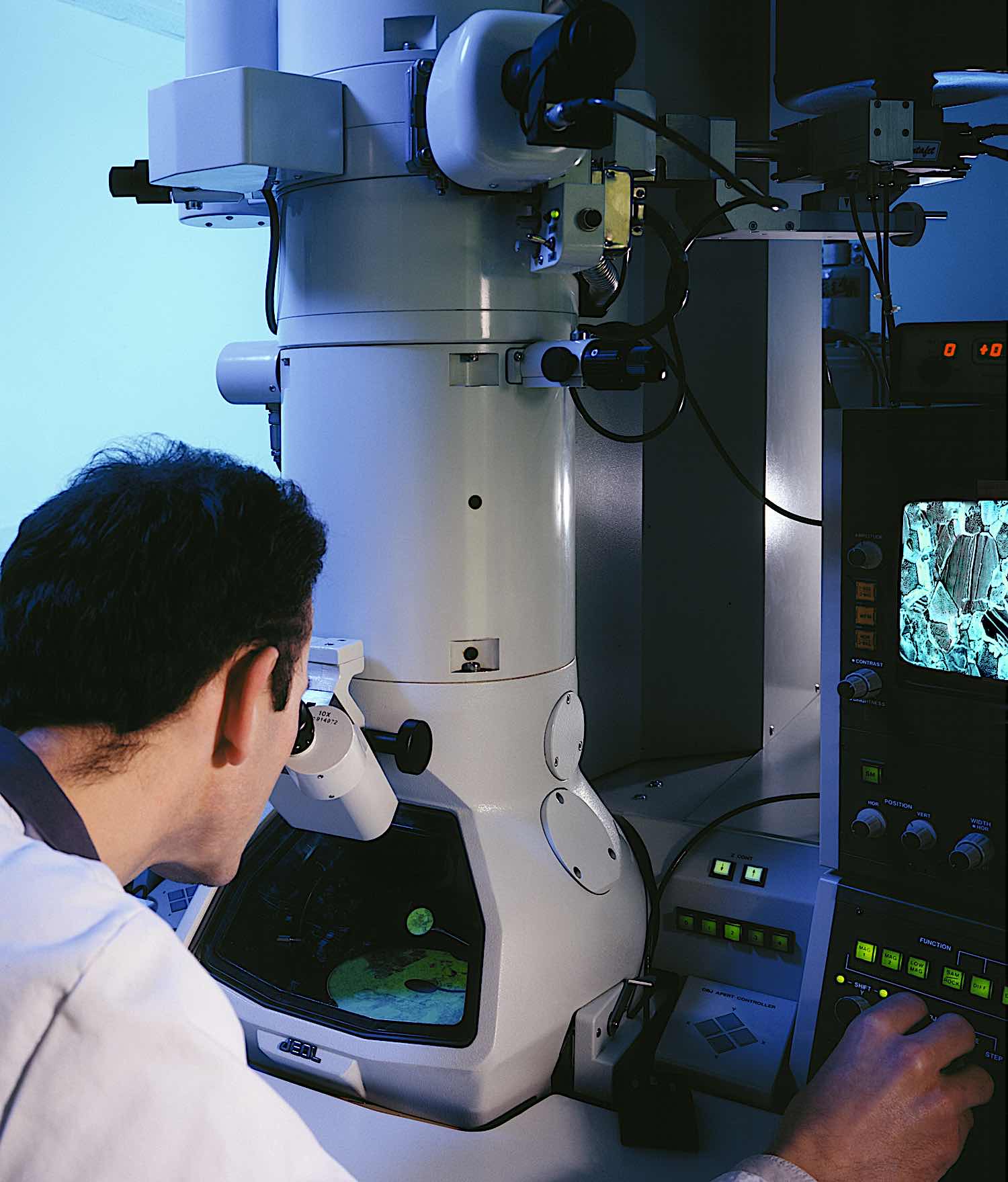
Microscopes enable humans to see things that are irreconcilably small, but all of them have a limit. This is known as the diffraction limit. Caused by the diffraction of light waves at the tiniest levels, it has prevented microscopes from seeing anything smaller than the light waves themselves.
Attempts at passing this barrier are numerous, and mostly used a technique that researchers have dubbed “superlensing”, which involves making custom lenses out of unique materials. Unfortunately, all these lenses gathered too much light to achieve the desired outcome.
Now however, a team of physicists from the University of Sydney claim to have discovered a means to surpass the diffraction limit by a factor of four times, and they achieved this in a way that no one else had tried before.

Superlensing without a superlens
Dr. Alessandro Tuniz from the School of Physics and University of Sydney Nano Institute is the lead author of this research, and he has said in a press release:
“We have now developed a practical way to implement superlensing, without a super lens. To do this, we placed our light probe far away from the object and collected both high- and low-resolution information. By measuring further away, the probe doesn’t interfere with the high-resolution data, a feature of previous methods.” [1]
Dr. Tuniz and his team overcame this by implementing the superlens capability as a post-processing step on a computer, after the measurement had actually been taken, thus producing a ‘truthful’ image of what was under the lens. This is done by selectively amplifying the evanescent (vanishing) light waves. [2]
In more detail, the probe gathered light “at terahertz frequency at millimetre wavelength, in the region of the spectrum between visible and microwave”, and this information was entered into a computer. Then, it was basically processed so that unwanted light could be removed, resulting in an image that goes beyond the diffraction limit. [3]

Previous attempts
Previous attempts at surpassing the diffraction limit focused on creating a superlens.
No matter how smooth and polished a conventional lens may be, there will always be a limited amount of detail that it can reproduce because of light’s tendency to diffract. This limit can be surpassed if only a way can be found to collect evanescent waves that exist close to an object’s surface, as these waves can resolve surface features which are far smaller than propagating waves. Unfortunately, these propagating waves decay too quickly for conventional lenses to capture.
Additionally, whatever material that scientists and researchers would use for their lenses, the lenses tended to collect too much light and become opaque.
This is why the team of researchers from The University of Sydney removed the superlens from the equation altogether and focused instead on computing power. They noted that previous attempts to select high-resolution information at a significantly closer range were unsuccessful as the most valuable data “decays exponentially with distance”, and so becomes overwhelmed by low-resolution data. The resulting image is then rendered useless. [4]
Co-author of the paper (published in the journal Nature), Professor Boris Kuhlmey has explained that the real key to their success was moving the probe further and further away and doing the superlensing in a computer. Doing so allowed them to preserve the integrity of their high-resolution data and use a post-observation technique to filter out low-resolution data. [5]
The applications of superlens technology
The research makes note of several possible applications of this new superlens technology, ranging from medicine and diagnostics to achievements that were previously unthought of.
Associate Professor Kuhlmey said of the tetrahertz range: “This is a very difficult frequency range to work with, but a very interesting one, because at this range we could obtain important information about biological samples, such as protein structure, hydration dynamics, or for use in cancer imaging.” [6]
Dr Tuniz said: “This technique is a first step in allowing high-resolution images while staying at a safe distance from the object without distorting what you see.
“Our technique could be used at other frequency ranges. We expect anyone performing high-resolution optical microscopy will find this technique of interest.” [7]
As Dr. Tuniz stated, while the virtual superlens technique was used and is suited to tetrahertz near-field photoconductive setups, it can easily be adapted for use in any near-field experiment measuring amplitude and phase. It could therefore allow for the increase in imaging resolution of near-field setups at any frequency. [8]
The capability to measure near fields without unsettling them could prove particularly useful in imaging fields that involve structures that are particularly sensitive to perturbations, such as photonic crystal defects, nanoresonators, and high-Q/topological resonances. [9]
Now that superlensing is here (in a sense), the quantity and quality of its application and uptake remain to be seen.
Sources
[1] – [5]
The Debrief – Breaking the diffraction limit by ‘superlensing’ without a superlens
[6] [7]
The University of Sydney – Superlensing without a superlens: microscopes boosted beyond limits
[8] [9]
Photonics Media – Virtual Superlens Exceeds Diffraction Limit Without Image Distortion


