
There are few parts of the human anatomy that are as delicate as the spinal cord. This essential but fragile part of the nervous system carries vital information between the brain and the rest of the body. Protected by the vertebrae of the spine, the spinal cord is difficult to access, which makes it challenging to study and treat conditions that affect it.
Researchers are studying less invasive techniques for exploring and treating the spinal cord. Now, innovative scientists have developed a new type of probe that could enable them to explore the spinal cord non-invasively.
The innovation
According to a recent press release from the University of California San Diego [1], their researchers, together with the Salk Institute for Biological Studies, have engineered a flexible neural probe that is one-fifth as wide as a human hair.
Jacob’s School demonstrates the ultra-thin flexible neural probe:
This multi-modal coaxial microprobe is designed to be inserted into the spinal cord with minimal disruption.
The size of this novel probe, 8 to 14 micrometers in diameter [2], enables it to examine dynamic areas of the nervous system, such as the spinal cord, while minimizing injury to the surrounding tissue.
According to Axel Nimmerjahn, co-senior author of the study and Associate Professor at the Salk Institute, a small, flexible probe can fit in between the vertebrae and interface with the neurons.

It can also bend as the spinal cord moves, making it ideal for studying the dynamics of the nervous system.
There are other small, flexible probes already available on the market.
What sets this new design apart is that it is the first probe to have a dual capacity for optical stimulation as well as for electrical recording of neural networks in such a small size.
Engineered Design
This innovative probe has been designed to have two channels:
- an electrical channel containing an ultra-thin polymer electrode
an optical channel that contains an also ultra-thin optical fiber
It wasn’t a straightforward task to put together these two channels of communication.
The researchers had to come up with a way to package them together in such a way that they could still function independently while being as small as possible.
This required insulation for each of the channels to stop them from interfering with each other while still fitting to the required minimal scale of 8 to 14 micrometers in diameter.
As well as the requirements of size, the probe had to be designed to be soft and flexible enough to follow the natural curves of the spine while still being stiff enough to be inserted with minimal damage. Biocompatibility was another design consideration, as the probe is inserted into living tissue.
In order to come up with a design that met all of these requirements, the team had to find the exact right combination of materials to use in building the probe and optimizing the electrical channel.
Biocompatibility
In order to come up with a usable design, the research team had to ensure the biocompatibility of their new probe. This meant that it couldn’t trigger an immune response when introduced to living tissue, as this would cause inflammation and damage to surrounding cells.
In an attempt to make it biocompatible, the new probe is coated with a thin layer of parylene. This is a common biomaterial that is used in a variety of medical devices, such as pacemakers and stents. [2]
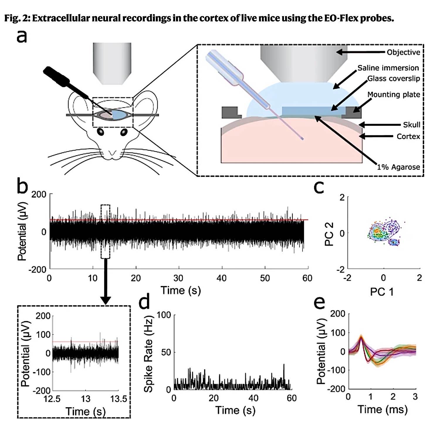
To ensure that their design was biocompatible, the team tested the probe on mice. They implanted the probes into the brains of live mice and monitored them over a month for any adverse effects.
Their findings were that even after such a long-term presence, the probe caused minimal inflammation in the brain tissues.
This led them to conclude that the device was compatible with biological tissue and, therefore, ideal for long-term use.
As Donald Sirbuly, professor of nanoengineering at the UC San Diego Jacobs School of Engineering and co-senior author of the study, pointed out, in order to allow chronic neural interfacing, the probe must be able to communicate with the neurons, but with the body barely knowing it is present.
Research findings
As Nimmerjahn remarked, we know very little about how the spinal cord processes information and how certain medical conditions can disrupt neural activity.
The team’s promising findings of their studies on mice, published in the journal Nature [2], show that the new probe is able to record neural activity with high fidelity while also stimulating neurons with light to cause certain physical reactions.
They found that they could make use of the probe’s optical channels to stimulate the neurons of the mice and get them to move their whiskers.
This proves that the new probe is not only able to record neural activity but also to influence it. This could have important implications for the treatment of conditions such as spinal cord injuries.
Applications in research and health technology
The tests carried out by the research team were proof of concept, and they hope to be able to use them in future studies.
The new probe will allow researchers to study the spinal cord in unprecedented detail to enable them to better understand the dynamics of the nervous system. The probe is highly customizable to fit specific requirements of length, diameter, or properties, so different research groups could use it to study different questions.
The probe could also be used in medicine, such as developing new treatments for neurological disorders or injuries.
Given the minimal inflammatory response caused by the probe, it could be used in a wide range of applications where neural activity needs to be monitored or influenced.
The UC San Diego and the Salk Institute have submitted a patent application for the new probe design. And it looks like, when it becomes more widely available, it could be a key to help to unlock some of the mysteries of the nervous system.
Notes
[1]: https://jacobsschool.ucsd.edu/news/release/3463
[2]: https://www.nature.com/articles/s41467-022-30275-x


