Many advancements have made in the field of science as of late, including a number of innovative discoveries regarding organ and cell research. From cell manipulation to lab-grown organs, scientists are working to prove that nothing is impossible.
Creating 3D Shapes from Living Tissue
Bioengineers from San Francisco recently published a report in the journal Developmental Cell, describing the creation of complex, folded shapes that could be replicated using living tissue. Some of these shapes included bowls, coils, and ripples, which were created by patterning mechanically active cells to thin layers of extracellular matrix fibers.
According to senior author Zev Gartner, “breaking the complexity of development down into simpler engineering principles” allowed scientists to “better understand, and ultimately control, the fundamental biology.”
“In this case, the intrinsic ability of mechanically active cells to promote changes in tissue shape is a fantastic chassis for building complex and functional synthetic tissues,” he said.
The goal in this research is to eventually create tissue “organoids,” or tiny lab-grown tissues that are typically grown from stem cells. These organoids are vital in research aimed at screening drugs and determining their effectiveness in treating a specific patient’s disease.
Alex Hughes, a postdoctoral fellow at UCSF, stated, “We’re beginning to see that it’s possible to break down natural developmental processes into engineering principles that we can then repurpose to build and understand tissues. It’s a totally new angle in tissue engineering.”
Gartner added, “It was astonishing to me about how well this idea worked and how simply the cells behave. This idea showed us that when we reveal robust developmental design principles, what we can do with them from an engineering perspective is only limited by our imagination. Alex was able to make living constructs that shape-shifted in ways that were very close to what our simple models predicted.”
The team is now working to determine how cells differentiate in response to the mechanical changes that occur during tissue folding in vivo. Inspiration for this research stems from stages of embryo development.
3D-Printed Artificial Organ Models
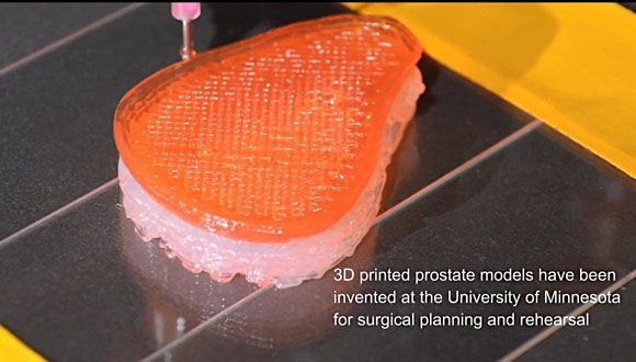
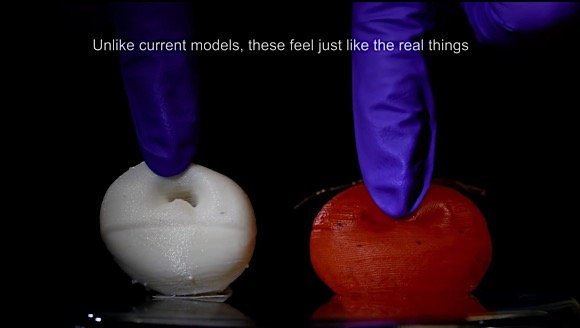
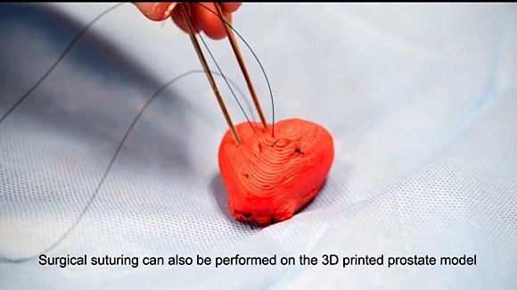
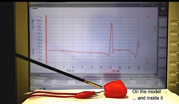
Researchers with the University of Minnesota recently created lifelike artificial organs capable of mimicking the exact anatomical structure and mechanical properties of their real-life counterparts. These models include integrated sensors that allow them to be used during practice surgeries, which result in improved surgical outcomes in patients around the world.
Lead researcher Michael McAlpine, an associate professor of mechanical engineering in the University of Minnesota’s College of Science and Engineering, discussed the research, saying, “We think these organ models could be game-changers for helping surgeons better plan and practice for surgery. We hope this will save lives by reducing medical errors during surgery.”
3D-printed organs are typically made using plastics and rubbers, which limits the application of these models in practice. Although certainly beneficial, they lack the realism that is necessary in accurate prediction and replication of the organ’s physical behaviour during surgery. Researchers developed these new models by taking MRI scans and tissues from patients’ prostates, testing them, and developing customized inks that can be altered to precisely match the unique properties of each patient’s prostate tissue.
According to lead author Kaiyan Qiu, a mechanical engineering postdoctoral researcher at the University of Minnesota, “The sensors could give surgeons real-time feedback on how much force they can use during surgery without damaging the tissue. This could change how surgeons think about personalized medicine and pre-operative practice.”
“if we could replicate the function of these tissues and organs, we might someday even be able to create bionic organs for transplants,” said McAlpine, describing what he calls the “Human X project,” which could lead to the eventual development of 3D-printed replacement organs for use in transplants.
Scientists Grow Hydrogel in the Same Manner as Biological Tissues
Scientists from Nanyang Technological University, Singapore and Carnegie Mellon University discovered a method for directing the growth of hydrogel to mimic biological tissues. Their findings were published in Proceedings of the National Academy of Sciences.
The paper suggests that this research opens the door for applications in areas that use hydrogel, such as tissue engineering and soft robotics. The group of scientists discovered that the manipulation of oxygen concentration allowed them to control the growth rate of hydrogels and create complex 3D shapes. Higher oxygen concentrations slowed the cross-linking of chemicals and inhibited growth.
The technique used by the research team differs from other methods of creating 3D structures through the addition or subtraction of layers of materials. This technique relies on continuous polymerization of monomers inside the porous hydrogel, which mimics the continuous growth of living systems, allowing researchers to study growth phenomena in those systems.
“Greater control of the growth and self-assembly of hydrogels into complex structures offers a range of possibilities in medical and robotics fields,” said Nanyang Technological University President Subra Suresh. “One field that stands to benefit is tissue engineering, where the goal is to replace damaged biological tissues, such as in knee repairs or in creating artificial livers.”
3D-Printing and Cryogenics Aim to Replicate Organs and Regenerate Damaged Parts
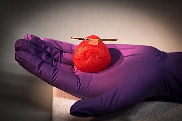
Researchers from Imperial College London developed a new technique for using cryogenics and 3D printing to create soft tissue structures that could be used in replicating organs as well as tissue regeneration. The process involves using dry ice as a cooling agent for hydrogel ink that is extruded from a 3D printer. Once thawed, it is soft like organic tissues, although it does not collapse under its own weight as in previous techniques.
The technique is currently being used on human skin, as researchers seed 3D-printed structures with dermal fibroblast cells, resulting in the regeneration of connective tissue in the skin. Research suggests that the technique has other potential uses as well, such as regenerating neuronal cells in the brain and spinal cord or creating replica body parts and organs for the purposes of medical training or for use in human transplants.
“At the moment, we have created structures a few centimetres in size, but ideally we’d like to create a replica of a whole organ using this technique,” said researcher Zhenchu Tan in a statement.
Biological research and innovative new discoveries such as these create new possibilities in the field of medicine. When it comes to organ and tissue development, these discoveries can result in thousands of lives being saved every year.
Sources:
University of California San Francisco


